Swiss Startup Connects 16 Human Mini-Brains to Create Low Energy ‘Biocomputer’
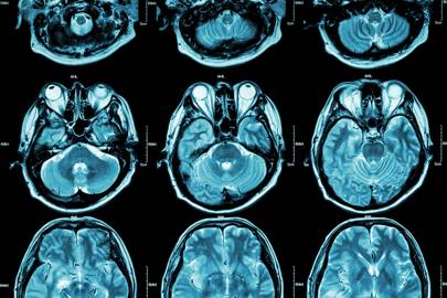
 The bioprocessor with eight electrodes attached to four arrays each housing a cluster of brain cells. (Jordan et al., Frontiers in Artificial Intelligence, 2024)
Computer scientists have for decades been vying to emulate the human brain, replicating its neural networks to build artificial intelligence (AI) with enhanced processing power.
But the more sophisticated those artificial neural networks become, the more powerful they get, and the more we rely on them, the more energy they consume. And sometimes nature’s original design is just better in some regards.
In the latest demonstration of nature’s efficiencies, a Swiss start-up company has just launched a ‘biocomputer’ that connects to living, pulsing brain cells and, according to its makers, uses far less energy than traditional, bit-based computers as a result.
Rather than merely integrating biological concepts into computing, FinalSpark’s online platform ‘taps’ into spherical clusters of lab-grown human brain cells called organoids. A total of 16 organoids are housed within four arrays that connect to eight electrodes each and a microfluidics system that supplies water and nutrients for the cells.
The approach, known as wetware computing, in this case harnesses researchers’ abilities to culture organoids in the lab, a fairly new technology that allows scientists to study what are essentially mini replicas of individual organs.
The rise in organoids as a popular research technique comes at a time when artificial neural networks, which underpin large language models such as Chat GPT, have also exploded in use and processing power.
FinalSpark claims that so-called bioprocessors like the brain-machine interface system they’re developing “consume a million times less power than traditional digital processors”.

While we don’t have any numbers on their specific system, its energy usage, or processing power, FinalSpark’s research team says that training a single large language model like GPT-3, a precursor to GPT-4, required 10 gigawatt hours or about 6,000 times the energy that one European citizen uses in a year.
Meanwhile, the human brain operates its 86 billion neurons using only a fraction of that energy: just 0.3 kilowatt hours per day.
Technology trends also indicate that the booming AI industry will consume 3.5 percent of global electricity by 2030. Already, the IT industry as a whole is responsible for around 2 percent of global CO2 emissions.
Clearly, it’s becoming increasingly necessary to find ways to make computing more energy efficient, and the synergies between brain cell networks and computing circuits are an obvious parallel to explore.
FinalSpark is not the first outfit to try connecting probes to biological systems, or attempt to reliably program neural networks so they perform specific input-output functions on command.
In 2023, researchers in the United States built a bioprocessor that connected computer hardware to brain organoids, and the system learned to recognize speech patterns.
“Over the past three years, the Neuroplatform was utilized with over 1,000 brain organoids, enabling the collection of more than 18 terabytes of data,” FinalSpark co-founder Fred Jordan and his colleagues write in their published paper, which has been peer-reviewed like other scientific studies.
While the end goal may be new, energy-efficient computing approaches, for now the system is being used to enable researchers to run lengthy experiments on brain organoids, just like its predecessors.
However, there are some improvements: The FinalSpark team says researchers can connect to its system remotely, and the mini-brains can be sustained for up to 100 days, their electrical activity measured around the clock.
“Currently in 2024, the system is freely available for research purposes, and numerous research groups have begun using it for their experiments,” Jordan and colleagues write.
“In the future, we plan to extend the capabilities of our platform to manage a broader range of experimental protocols relevant to wetware computing,” such as injecting molecules and drugs into organoids for testing, the team concludes.
Whichever way this goes, aiding computing or organoid research, it’ll be exciting to see what researchers can achieve.
The study has been published in Frontiers in Artificial Intelligence.
from: https://www.sciencealert.com/swiss-startup-connects-16-human-mini-brains-to-create-low-energy-biocomputer